
Eye Testing Technology at RJK Optometry, Coffs Harbour
At RJK Optometry we have invested in modern diagnostic technology to help detect eye conditions early.
The diagnostic equipment we use in Coffs Harbour
RJK Optometry, an independent optometry practice at 14 Arthur Street, Coffs Harbour, uses Optos widefield retinal imaging, OCT (optical coherence tomography), corneal topography, the Rodenstock DNEye scanner and ImpressionIST 3, and dry eye assessment equipment, together with the M22 Optima IPL and Rexon-Eye treatment devices. Here is what each one does.
- Optos Daytona widefield retinal imaging: captures an image of up to 200° of the retina (the light-sensitive layer at the back of the eye) in a single scan. It helps us look for signs of conditions such as glaucoma, macular degeneration, diabetic eye disease and ocular melanoma, often without dilating drops, although dilation may still be recommended for some people.
- OCT (optical coherence tomography): takes cross-sectional scans that measure the layers of the retina and the optic nerve fibres. It is used to help assess and monitor glaucoma and macular conditions over time.
- Corneal topography: maps the shape of the front surface of the eye (the cornea). We use it when fitting contact lenses and Ortho-K, and to check whether Ortho-K suits you.
- Rodenstock DNEye scanner: takes thousands of measurements of how your eye focuses light, which are used to personalise Rodenstock lenses for you.
- Rodenstock ImpressionIST 3: a camera-based system that measures how your chosen frame sits on your face, in 3D, to an accuracy of 0.1mm.
- Dry eye assessment equipment: used to look at the tear film and the oil glands in the eyelids (the Meibomian glands), so we can work out what is causing dry eye symptoms.
- Eye pressure and visual field testing: as part of a comprehensive eye test, we measure the pressure inside your eye and, where needed, test your peripheral (side) vision. Both help assess the risk of glaucoma.
- M22 Optima IPL (Lumenis): delivers controlled pulses of light around the eyelids. It is a TGA-approved treatment option for Meibomian Gland Dysfunction.
- Rexon-Eye: uses gentle electrical stimulation (Quantum Molecular Resonance), which aims to improve how the tear glands work. We’ll discuss whether either dry eye treatment suits you.
DNEYE scanner
Our equipment includes the Rodenstock DNEye scanner.
The DNEYE scanner is used to capture more than 7,000 data points from the eye, which gives us detailed information about how your individual eye focuses, which we use to personalise your lenses. Many patients notice better contrast, though results vary.


Rodenstock ImpressionIST 3
Optical lenses need to be fitted precisely for clear vision.
Even high-quality lenses need to be precisely fitted to your frame. This used to be done by hand. We use digital measurement technology to determine the precise centration of your lenses in your frame, as you wear it. The Rodenstock ImpressionIST 3 is a computerised measuring system which draws on German expertise in engineering. With its unique dual camera technology, the ImpressionIST 3 calculates the position of all points of your frame and your eye in 3D space to an accuracy of 0.1mm.
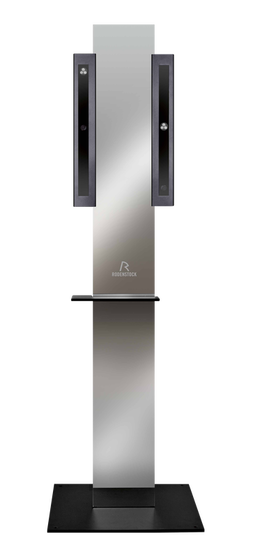
Every frame and every face is different.
- Five main parameters are required in order to produce a highly customised lens:
- The distance from your pupil to the back of the lens (vertex distance)
- The angle the lens sits at in front of your eyes (pantoscopic tilt)
- The curvature of the frame (frame wrap angle)
- The height of your pupil in the frame as it is worn
- The horizontal centration of your pupil in the frame
Once these measurements are taken, Rodenstock incorporates them to the calculations in order to optimise the optical characteristics of your lens. The ImpressionIST uploads the data directly to the optical lab in Germany where your lenses are manufactured. With German precision, the lenses are digitally cut in a process known as freeform production.
Your lenses are designed for you, to help give clear near and distance vision with less distortion.
Optos Daytona Widefield retinal Scan
The Ultra Wide Digital Retinal Scan gives us a 200° range within a single scan capturing highly detailed images of your retina.
The scan helps us identify any blood vessel abnormalities that could indicate early signs of disease. We recommend an annual retinal scan for all adult patients.
The Digital Retinal Scan aids in the early diagnosis of glaucoma, macular degeneration, ocular melanoma and diabetic eye disease.
OCT Optical Coherence Tomography
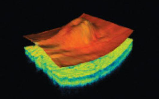
Optical coherence tomography (OCT) provides a detailed way of examining the structure of the retina.
In certain conditions different layers of the retina are affected. The bottom most layeris usually first affected in macular degeneration. The retinal nerve fibre layer is thinned in glaucoma.
An OCT allows detailed measurement of these layers and can be used as an additional tool to track disease.


